FOLLOW-UP
"The young intern has no
experience in dealing with
the submission of specimens
for histopathology," I
explained to the Laboratory
girl who phoned me to tell
me that the 7 testicular
specimens must be
correctly labelled as
"right" and "left" testicle
when I queried the report's
findings. My
histopathological samples
are sent to a commercial
laboratory that specialises
in this subject.
How come there was a
blunder? It was my policy of
letting interns be more
hands-on.
I had instructed my
assistant, Mr Saw, to let
vet interns have some
hands-on experience rather
than being an observer. In
that way, they will learn
from experience and
mistakes. We all learn every
day as we can't be perfect
people.
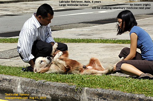
Ms Lai, the intern, would be
studying veterinary medicine
next year and so we gave her
the task of submission and
filling out the forms. She
had been with us for around
2 weeks and had seen
submission of blood samples.
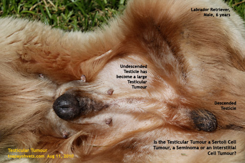
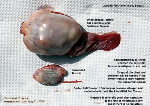
Well, the undescended
testicle of this 6-year-old
Retriever was enormous (half
the size of a tennis ball)
and would not fit into the
usual container we used to
submit samples for
histopathology. So, I
advised cutting it into 3
smaller pieces for the
larger testicle (left) and 4
smaller pieces for the
descended atrophied testicle
(right).
I ought to have advised
getting a bigger container.
I did that eventually. Next
time I asked my assistant to
check before submission of
the samples. Usually we
submit one sample and it
fits nicely inside the
Lab-provided bottles.
Everybody learns from being
hands-on. Young interns
can't be spoon-fed all the
time and will make some
mistakes. It is part of
growing up in life.
I was fortunate to have
worked in the Vet Diagnostic
Lab at Kampong Java Road
(now the new Kandang Kerbau
Hospital) as a new graduate
in 1977 and so knew a bit
about the work of
histopathology. What had
happened was that the intern
put 7 pieces 1 big bottle
and submitted the bottle and
form without separation of
the left and right testicle.
After receiving the
histopathology reports, I
phoned to clarify that there
were two testicles submitted
as the report had presumed I
had submitted 7 pieces of
one testicle. The report was
then corrected as follows:
Specimens 1-4 are left
undescended testicle.
Specimen 5-7 are the
atrophied descended right
testicle. The histopathology
results are as follows:
Takes a long time to
pee. Painful enlarged
prostrate when digital
examined via rectum
during light
anaesthesia. Could
this be purulent
prostatitis as
mentioned in:
www.vet.uga.edu/vpp/clerk_anat
/sabatino/index.php.
Hyperpigmentation in
the preputial area
Follow up 7 days
later: Owner is
happy. "No more
limping now. We
thought he had hip
dysplasia."
I asked" Was the dog
attracting other dogs
during exercise
outdoors?" The
owner recalled that
many dogs wanted to
make friends with him
(the effects of
hyperestrogenism).
Left testis -
Sertoli cell tumour
(see comments below).
Right testis - Atrophy
of the contra-lateral
scrotal testicle
resulting in
aspermatogenesis.
Comments from the
Histopathology Lab:
1. The left testis is
extensively replaced by a
tumour which is composed of
closely packed tubules,
trabeculae and nests of
elongated spindly cells. A
second interstitial (Leydig)
cell tumour component cannot
be excluded. Most primary
testicular neoplasm in dogs
are benign. The rare
malignant Sertoli cell
tumours have no good
cytological or histological
markers of malignancy. The
vet needs to identify
metastases in lymphatics,
spermatic cord, lymph node
or distant sites. Please
correlate with clinical
features.
Dr Sing's comments:
The left testis will likely
be a mixed tumour with
Sertoli and interstitial
cells involved). Much more
details of the types of
testicular tumour in the dog
are in an excellent report
at:
www.vet.uga.edu/vpp/clerk_anat/sabatino/index.php.
2. In the right
testis, the parenchyma shows
closely packed seminiferous
tubes composed solely of
Sertoli cells without
spermatogonia, spermatocytes
or spermatids. This is
common in cryptorchid
(undescended) testis. There
is no tumour
involvement.
CLINICAL FEATURES OF
INTEREST TO VET
UNDERGRADUATES
1. As the histopathologist
is an independent service
provider, he or she had not
seen the real dog. The right
testis was assumed to be an
undescended or cryptorchid
testis. It was a descended
testicle, much shrunken.
2. Hyperestrogenism. The
reason the descended right
testes was atrophied and had
no sperm production was due
to the excessive production
of estrogen by the large
left testicular tumour.
Estrogen is anti-androgenic
and therefore cause the
atrophy of the scrotal right
scrotal testes.
3. Blood test results as
shown below indicated a
thrombocytopenia which can
be a cause of death in the
dog if the tumour had not
been detected early and
removed.
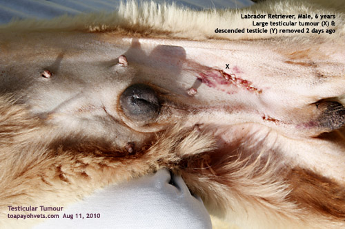
Golden
Retriever, Male, 6
years. Left
undescended testicular
tumour and atrophied
right scrotal testicle
removed 5 days ago.
Greyish-white, large
sized and
multi-nodular
suggestive of a
Sertoli cell tumour.
The owner asked about
post-op management.
This depended on blood
test results.
Blood tests:
1. No disorders of
liver and kidney
function. Glucose is
normal.
2. Haematology: Low
haemoglobin and red
cell count. Low PCV.
Very low platelet
count. Platelet
clumping noted. (This
indicated bone marrow
depression). Effects
are well written in:
www.vet.uga.edu/vpp/clerk_anat/sabatino/index.php.
This indicated a
severe bacterial
infection of the
bladder and prostate
(painful and enlarged
during rectal
palpation and presence
of "epithelial
cells"). The infection
is localised to the
urinary tract as the
total blood White Cell
Count was OK. The dog
had been given IV Vit
K1 in drip earlier and
appeared much more
energetic the next
day. Diagnosis would
be urinary tract
infection (UTI).
But the cause was the
testicular neoplasm
(hyperestrogenism
leading to bone marrow
depression and
prostatitis with
consequent UTI).
Advices to owner:
1. Had been fed meat,
rice 1X/day in the
past. Very thin.
Increase feed to
2x/day.
2. Good quality
premium dog food dry
to be added. 1 egg/day
for 14 days.
3. Antibiotics for
next 14 - 20 days. UTI
+ prostatitis + bone
marrow depression.
4. Review in 14 days.
Some 10 days later,
the owner said that
the dog was normal and
active. "I thought he
had hip dysplasia," he
told me over the
phone. "He was walking
with a limp."
I note that the "Summary"
stated that "the Sertoli
cell tumour is the only
known testicular tumour that
commonly produces hormones
with clinical effects." Some
vet reports I read on the
internet claim that estrogen
is produced by two of the 3
common testicular tumours,
namely the Sertoli cell
tumours and Seminomas.
In the "Introduction", there
was this statement that
"testicular neoplasms other
than Sertoli cell tumours
are rarely hormonal
productive. Testicular
neoplasms are often
mixed-origin especially in
cryptorchid testes". In this
case, the histopathologist
indicated that the
undescended testicle could
be a mixed type with Sertoli
cell tumour and interstitial
(Leydig) cell tumour.
Cryptorchidism
(undescended
testicles)
in the
dog can be unilateral
or bilateral
Note that the
above-mentioned
article
said that testicular
neoplasm in the dog is
presented early as 6
years of age (as in this
case-study) but the median
age is 10 years (photo
of one of my cases,
right).
In 2010, many younger
dog owners do not wish
to have their male
dogs neutered as they
perceive the surgery
to be cruel. However,
undescended testicles
may develop testicular
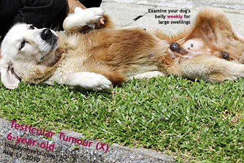
tumours. Weekly check
up of the dog's belly
will detect their
presence
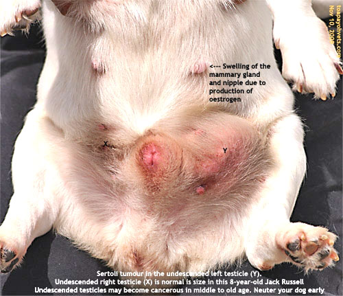
Testicular Tumour in an undescended testicle of an 8-year-old
Jack Russell.
The contra-lateral
testicle is atrophied.
Check the belly of
your older dog every
week if you do not
wish to have your male
dog neutered.
Testicular tumours are
seldom malignant but
they do cause death
due to bone marrow
depression and
thrombocytopenia and
consequent
overwhelming bacterial
infections.